May 28, 2020
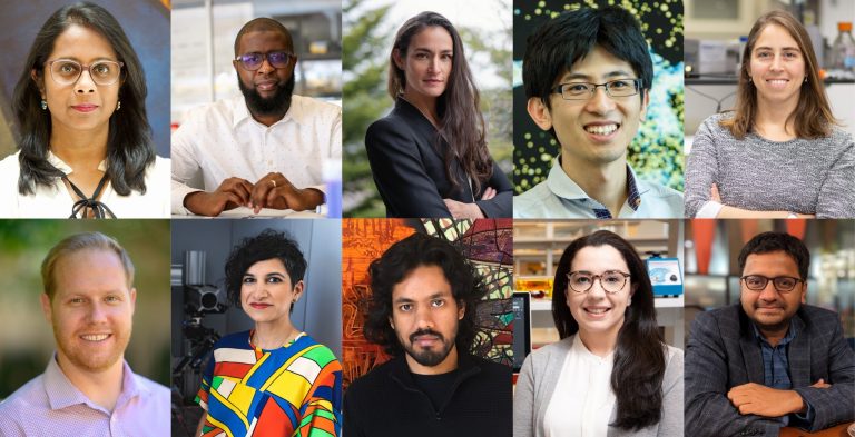
The Board of Directors of The McKnight Endowment Fund for Neuroscience is pleased to announce it has selected six neuroscientists to receive the 2020 McKnight Scholar Award.
The McKnight Scholar Awards are granted to young scientists who are in the early stages of establishing their own independent laboratories and research careers and who have demonstrated a commitment to neuroscience. “This year’s Scholars exemplify the power of modern neuroscience to elucidate the biology of the brain and mind,” says Kelsey C. Martin, M.D, Ph.D, chair of the awards committee and dean of the David Geffen School of Medicine at UCLA. Since the award was introduced in 1977, this prestigious early-career award has funded more than 240 innovative investigators and spurred hundreds of breakthrough discoveries.
“Leveraging an array of methodological approaches in diverse model organisms, the 2020 McKnight Scholars are advancing the neuroscience of gut-brain interactions and parent-infant bonding, deciphering the computational logic of motor planning in the cerebellum and the gene regulatory logic of inhibition in the cortex, identifying and characterizing novel chloride channels in neurons, and using structure-based approaches to develop new therapeutics that target specific serotonin receptors,” says Martin. “On behalf of the entire committee, I would like to thank all the applicants for this year’s McKnight Scholar Awards for their innovative scholarship and contributions to neuroscience.”
Each of the following six McKnight Scholar Award recipients will receive $75,000 per year for three years. They are:
Steven Flavell, Ph.D.
Massachusetts Institute of Technology – Cambridge, MA
Elucidating Fundamental Mechanisms of Gut-Brain Signaling in C. elegans
Studying how gut bacteria influence brain activity and behavior.
Nuo Li, Ph.D.
Baylor College of Medicine – Houston, TX
Cerebellar Computations During Motor Planning
Researching the process by which different parts of the brain, including the cerebellum, coordinate to plan physical motion.
Lauren O’Connell, Ph.D.
Stanford University – Stanford, CA
Neuronal Basis of Parental Engrams in the Infant Brain
Studying what happens in the brains of infant animals during parental bonding, and the effects this neuronal process has on future decision-making and well-being in adulthood.
Zhaozhu Qiu, Ph.D.
Johns Hopkins University – Baltimore, MD
Discovering Molecular Identity and Function of Novel Chloride Channels in the Nervous System
Research into the genes underlying diverse chloride channels, and their role in regulating neuronal excitability and synaptic plasticity.
Maria Antonietta Tosches, Ph.D.
Columbia University – New York, NY
The Evolution of Gene Modules and Circuit Motifs for Cortical Inhibition
Exploring the evolution of neural circuits by studying ancient neuron types in animals with simple brains to infer fundamental principles of brain organization and function.
Daniel Wacker, Ph.D.
Icahn School of Medicine at Mount Sinai – New York, NY
Accelerating Drug Discovery for Cognitive Disorders Through Structural Studies of a Serotonin Receptor
Determining the structure of a specific serotonin receptor linked to cognition, and using that structure to identify compounds that may bind to the receptor in a specific way to advance the discovery of drug therapies.
There were 58 applicants for this year’s McKnight Scholar Awards, representing the best young neuroscience faculty in the country. Faculty are only eligible for the award during their first four years in a full-time faculty position. In addition to Martin, the Scholar Awards selection committee included Dora Angelaki, Ph.D., New York University; Gordon Fishell, Ph.D., Harvard University; Loren Frank, Ph.D., University of California, San Francisco; Mark Goldman, Ph.D., University of California, Davis; Richard Mooney, Ph.D., Duke University School of Medicine; Amita Sehgal, Ph.D., University of Pennsylvania Medical School; and Michael Shadlen, M.D., Ph.D., Columbia University.
Applications for next year’s awards will be available in August and are due on January 4, 2021. For more information about McKnight’s neuroscience awards programs, please visit the Endowment Fund’s website at https://www.mcknight.org/programs/the-mcknight-endowment-fund-for-neuroscience
About The McKnight Endowment Fund for Neuroscience
The McKnight Endowment Fund for Neuroscience is an independent organization funded solely by The McKnight Foundation of Minneapolis, Minnesota, and led by a board of prominent neuroscientists from around the country. The McKnight Foundation has supported neuroscience research since 1977. The Foundation established the Endowment Fund in 1986 to carry out one of the intentions of founder William L. McKnight (1887-1979). One of the early leaders of the 3M Company, he had a personal interest in memory and brain diseases and wanted part of his legacy used to help find cures. The Endowment Fund makes three types of awards each year. In addition to the McKnight Scholar Awards, they are the McKnight Technological Innovations in Neuroscience Awards, providing seed money to develop technical inventions to enhance brain research; and the McKnight Neurobiology of Brain Disorders Awards, for scientists working to apply the knowledge achieved through translational and clinical research to human brain disorders.
2020 McKnight Scholar Awards
Steven Flavell, Ph.D. Assistant Professor, The Picower Institute for Learning and Memory, Massachusetts Institute of Technology, Cambridge, MA
Elucidating Fundamental Mechanisms of Gut-Brain Signaling in C. elegans
In recent years, there has been increased interest in the microbiome of the gut – the mix of bacteria living in the digestive tract – and its impact on overall health. Dr. Flavell will conduct a series of experiments to answer fundamental questions about how the gut and brain interact, how the presence of certain bacteria activates neurons and how this influences an animal’s behavior. This research could open new lines of inquiry into the human microbiome and how it influences human health and disease, including neurological and psychiatric disorders.
Little is understood about how the gut and brain interact mechanistically – which neurons are activated by the presence of bacteria? What are they detecting? What signals do they send and where? And how does the brain process these signals and turn them into behavior? Dr. Flavell’s research will build on discoveries his lab has made studying the C. elegans worm, whose simple and well-defined nervous system can generate relatively complex behaviors that are easily studied in the lab.
Dr. Flavell and his team have identified a specific type of enteric neuron (neurons lining the gut) that is only active while C. elegans feed on bacteria. His experiments will identify the bacterial signals that activate the neurons, examine the roles of other neurons in gut-brain signaling, and examine how feedback from the brain influences the detection of gut bacteria. For example, the enteric neurons of C. elegans signal to the brain when they detect bacteria, so that the worm can slow down and forage. The experiments will identify the nuances of this process, such as how signaling and behavior change when the worm is full or encounters different types of bacteria, and what happens when the activity of the enteric neurons is disrupted. Understanding these core processes may help future research unlock how gut bacteria in humans is linked to complex behavioral and neurological states.
Nuo Li, Ph.D., Assistant Professor of Neuroscience, Baylor College of Medicine, Houston, TX
Cerebellar Computations during Motor Planning
Timing is everything when it comes to moving muscles in a planned way. Dr. Li’s research uses a mouse model to explore in greater detail than previous studies what the brain is doing during the time between the plan and the movement. The old, simplified view of the brain used to envision the frontal cortex, where reasoning takes place, as the control center, and the cerebellum, an ancient part of the brain, as a tool to send signals to muscles. That view has become more nuanced, with researchers postulating that multiple parts of the brain are involved in thinking and planning.
Dr. Li’s lab has revealed that the anterior lateral motor cortex (ALM, a specific part of the mouse frontal cortex) and the cerebellum are locked in a loop while the mouse is planning an action. Still unknown is exactly what information is being passed back and forth, but it is distinct from the signal that actually drives the muscles. If the connection is disrupted even for an instant during planning, the movement will be made incorrectly. On the other hand, the brain can also use that time to convert feedback into improved planning for a subsequent movement, the way a basketball player adjusts after observing a miss.
Dr. Li’s experiments will uncover the role of the cerebellum in motor planning and define the anatomical structures that link it and the ALM. He will map the cerebellar cortex and find out which populations of a special type of cell used in cerebellar computation, called Purkinje cells, are activated by the ALM in motor planning, and what signals they send back and forth while planning. A second aim will explore what kind of computation the cerebellum is engaged in. The experiment uses mice who are trained to make a specific action some time after they observe a signal. By observing what parts of the brain activate during that anticipatory time when the animal is not moving but is preparing to move, and then by perturbing that process, Li will learn more about these sophisticated, fundamental brain processes.
Lauren O’Connell, Ph.D., Assistant Professor of Biology, Stanford University, Stanford, CA
Neuronal Basis of Parental Engrams in the Infant Brain
Parent/infant bonding is critical for the well-being of entire communities, in humans as well as animals. Not only does it support physical health, it also impacts the behaviors and choices of individuals as they reach adulthood. Dr. O’Connell’s work will help identify how memories are formed in infancy as part of the bonding process, will trace those memory imprints to identify how they affect future decision-making, and will explore the neurological impact of disrupted bonding.
This project uses a poison frog model, chosen because of its parent/infant bonding behavior seen in food provisioning by the parents. An additional benefit to the poison frog model is the frog’s physiology, which allows for clear observation of neural behavior. Bonding behavior is ancient and appears in brain regions that have been relatively conserved from amphibians to mammals. While there has been research examining the impact of bonding from a parental perspective, little is understood about how it occurs in infants or its neurological impact.
In the frogs O’Connell is studying, bonding behavior includes a begging display by the tadpoles, which leads the parent to provide unfertilized eggs for food. Receiving that food and care leads the tadpole to imprint on the parent, which in turn affects the tadpole’s future choice of mate: it will prefer mates that look like the caregiver. O’Connell has identified neuronal markers that are enriched in tadpoles that beg for food, and found that these neurons are analogous to those implicated in a range of neurological issues related to learning and social behavior in humans. Her research will explore the neuronal architecture involved in infant recognition and bonding with caregivers, as well as brain activity when making mate choices later in life, to see how neuronal activity in each process is related in normal conditions and when bonding is disrupted.
Zhaozhu Qiu, Ph.D., Assistant Professor of Physiology and Neuroscience, Johns Hopkins University, Baltimore, MD
Discovering Molecular Identity and Function of Novel Chloride Channels in the Nervous System
Ion channels are the cornerstones for the brain to maintain its normal function. They control neuronal membrane potential and excitability as well as synaptic transmission and plasticity. They are involved in many neurological and psychiatric disorders, and are thus major drug targets. Much of the research has been focused on ion channels conducting positively-charged ions, such as sodium, potassium and calcium. However, the function of ion channels allowing the passing of chloride, the most abundant negatively-charged ion, remains poorly understood.
One of the major challenges is the unknown identity of the genes encoding some of the diverse chloride channels. By performing high-throughput genomics screens, Dr. Qiu and his research team have identified two new families of chloride channels, activated by cell volume increase and acidic pH, respectively. Using a combination of electrophysiological, biochemical, imaging and behavioral techniques, Dr. Qiu’s research aims to investigate the neurological function of these new ion channels with a focus on neuron-glia interactions, synaptic plasticity, and learning and memory.
Dr. Qiu will extend this approach to other mysterious chloride channels in the brain. He also plans to develop new methods and tools to measure and manipulate chloride concentration at cellular and subcellular levels in the live cells and animals, which currently presents a major technical hurdle in this field. His research will provide key insights into how chloride is regulated in the nervous system. It may lead to novel therapeutics for neurological diseases associated with chloride dysregulation.
Maria Antonietta Tosches, Ph.D., Assistant Professor, Columbia University, New York, NY
The Evolution of Gene Modules and Circuit Motifs for Cortical Inhibition
It can be tempting to view the brain as an engineering feat, optimally designed to execute its complex functions. In reality, modern brains were shaped by a long evolutionary history, where at any point of an evolutionary challenge, existing components were repurposed, multiplied and diversified. Dr. Tosches is conducting research to understand these processes and figure out what fundamental neural systems have been conserved in vertebrate animals separated by hundreds of millions of years of evolution.
To that end, Dr. Tosches is exploring the evolutionary history of GABAergic neurons, which play an important inhibitory role in the mammalian central nervous system. Her previous experiments have found the GABAergic neurons of reptiles and mammals are genetically similar, indicating that these neuron types existed already in vertebrate ancestors; they also share gene modules associated with specific neuronal functions in both types of brains. In Tosches’ new research, she will determine if these same neuron types are found in the simple brain of salamanders.
The research involves sequencing tens of thousands of individual cells from these salamanders and comparing the GABAergic cell types found to those in mice and turtles, to build a unified taxonomy of these neurons in tetrapods. The next step is to compare their gene modules to understand the genetic mechanisms that have given rise to GABAergic neuron subtypes. In a second aim, Tosches and her team will record the activity of salamander GABAergic neurons with in vivo imaging during behavioral experiments, tracking the activity of these neurons when presented with stimuli. This work will introduce a completely new animal model to circuit neuroscience, adding to our understanding of how the brain works at a fundamental level.
Daniel Wacker, Ph.D., Assistant Professor, Icahn School of Medicine at Mount Sinai, New York, NY
Accelerating Drug Discovery for Cognitive Disorders through Structural Studies of a Serotonin Receptor
Discovering drugs to address neurological and cognitive disorders is a complex and time-consuming process. Many drugs target dopamine receptors which are linked to addiction, and some drugs are imprecise and create potentially dangerous side effects. Additionally, some disorders (Alzheimer’s is a prominent example) have no drug treatments at all. Dr. Wacker proposes a novel approach to drug discovery that focuses in on a specific serotonin receptor (which doesn’t carry the same risks as activating the dopamine system), carefully mapping that receptor’s structure at a molecular scale, and seeking out compounds that will bind to that receptor in a specific way.
The receptor, known as 5-HT7R, was discovered in the mid-1990s and is one of 12 known serotonin receptors. It has been identified as a promising target for therapies for cognitive disorders, but little is known about it. Dr. Wacker proposes to conduct a structural study of the receptor using X-ray crystallography on purified samples of the receptor. He will examine how drugs bind to the receptor and introduce mutations to the structure to see how that affects the binding and interaction. The goal is to find compounds that will activate just this one receptor in a specific way.
To find these possible drugs, Wacker’s team will then conduct a computerized search of hundreds of millions of compounds, comparing their 3D structure with the 3D model of the receptor for those most likely to “fit.” Top prospects will be inspected more closely, and a few especially promising candidates will be tested in the lab. Compared with a traditional process of drug trials, which can take years, even decades, this computerized process offers the opportunity to essentially pre-screen drugs based on their structure, and speed their development.


