August 1, 2022
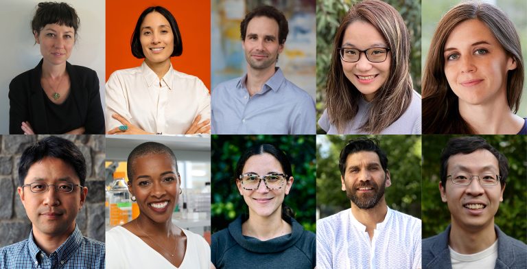
The McKnight Endowment Fund for Neuroscience (MEFN) announced the three recipients of $600,000 in grant funding through the 2022 McKnight Technological Innovations in Neuroscience Awards, recognizing these projects for their ability to fundamentally change the way neuroscience research is conducted. Each of the projects will receive a total of $200,000 over the next two years, advancing the development of these groundbreaking technologies used to map, monitor, and model brain function. The 2022 awardees and their projects:
- Andre Berndt, PhD, of the University of Washington, is developing a system to create and scan very large numbers of optogenetic biosensors very quickly, so that researchers can identify and refine these biosensors more precisely for their experiments. Current technology and resource constraints limit researchers to exploring mere dozens or hundreds of biosensors, and the small sample size means they can’t be sure they found the best option. With the ability to create and screen tens of thousands, their options will expand exponentially.
- Ruixuan Gao, Ph.D., of the University of Illinois Chicago, is chemically engineering a new type of hydrogel to use in a new practice of expansion microscopy – essentially expanding tissue samples and their component cells to many times their original size to make them easier to study. His new “tetra-gel” and specialized molecules that anchor the sample to the gel will allow it to expand with high fidelity and remain stable so that the molecular profile of brain tissue can be better captured.
- Mirna Mihovilovic Skanata, Ph.D., of Syracuse University, is developing a new, high-precision application for two-photon microscopy that will allow researchers to precisely track and optically manipulate neural activity over a large area in freely-behaving, freely-moving larval fruit flies. The system is totally non-invasive, using an algorithm to adjust for the motion of the larvae and keep track of multiple individual cells simultaneously by calculating and correcting for the motion and deformation brain as the animal moves.
Learn more about each of these research projects below.
About the Technological Innovations in Neuroscience Awards
Since the McKnight Technological Innovations in Neuroscience Award was established in 1999, the MEFN has contributed more than $16 million to innovative technologies for neuroscience through this award mechanism. The MEFN is especially interested in work that takes new and novel approaches to advancing the ability to manipulate and analyze brain function. Technologies developed with McKnight support must ultimately be made available to other scientists.
“Again, it has been a thrill to see the ingenuity that our applicants are bringing to new neurotechnologies,” said Markus Meister, Ph.D., chair of the awards committee and the Anne P. and Benjamin F. Biaggini professor of biological sciences at Caltech. “Our awards span a broad range, from new biosensors for signaling molecules to clever methods that expand neural tissue before high-resolution microscopy.”
This year’s selection committee also included Adrienne Fairhall, Timothy Holy, Loren Looger, Mala Murthy, Alice Ting, and Hongkui Zeng, who chose this year’s Technological Innovations in Neuroscience Awards from a highly competitive pool of 90 applicants.
For more information about the awards, please visit our website.
2022 McKnight Technological Innovations in Neuroscience Awards
Andre Berndt, PhD, Assistant Professor, Department of Bioengineering, University of Washington
Massively parallel, high throughput engineering of optogenetic biosensors for neuronal signaling
Florescent, genetically encoded proteins have revolutionized the study of brain cells and neural circuits. By literally lighting up in the presence of specific neural activity, which then can be recorded by microscopes and light fibers in living brains, this tool has unlocked many mysteries and allowed researchers to visualize brain activity and neural pathways. But there has been a bottleneck: Developing and identifying the best sensor for each experiment. These encoded proteins need to react in the presence of only specific stimuli, in some cases might need to be highly sensitive, in other cases may need to fluoresce for a longer period of time, or an experiment may need two sensors to see how multiple neurotransmitters interact.
In the past, each sensor had to be genetically modified, produced, and tested individually. Perhaps only a few dozen or hundred could be compared, and researchers chose the best option from a small sample – not knowing if there was a better, more precise option available. Dr. Berndt has developed a process for developing and testing very large numbers of optogenetic biosensors simultaneously, aiming for screening more than 10,000 per day and building a massive library of biosensors that can give researchers access to precisely engineered proteins they can use to run ever-more specific experiments.
The technology uses rapid genetic engineering to create large numbers of variants of a biosensor, then places individual variants into a microwell array. The sensors are exposed to neuropeptides – currently, Dr. Berndt is focusing on ligand-specific opioid sensors – and optical sensors then read the microarray, detecting the brightness and other variables of each variant, and selecting the best options for further testing. Over the course of 2 years, some 750,000 biosensors will be tested and the process for their screening refined, advancing research into opioid actions in the brain and providing a versatile approach other researchers can use for their experiments.
Ruixuan Gao, Ph.D., Assistant Professor, Department of Chemistry and Department of Biological Sciences, University of Illinois Chicago
Sub-10 nm spatial profiling of synaptic proteins and RNA transcripts with high-isotropy expansion microscopy using a highly homogenous hydrogel constructed from tetrahedron-like monomers
To examine things that are very small – like the neurons and their synapses in the brain – researchers use powerful microscopes. But there is another approach that can yield impressive results: literally expanding a tissue sample and the cells within it by use of a special swellable hydrogel through a process called expansion microscopy. The hydrogel binds to different molecular components of cells and expands, ideally holding all the component parts in the same relative position to one another, creating a larger and more accessible sample to study – in principle, similar to writing on a balloon, then inflating it.
However, the current hydrogels used for this process have some drawbacks when it comes to studying minute structures in the brain. The margin of error in holding the relative position of molecules isn’t as precise as desired. New gel that potentially overcomes this issue reacts poorly to the heat used in denaturing and treating tissue samples. And it can limit the use of fluorescing biomarkers. Dr. Gao aims to improve the technology by developing a new type of “tetra-gel”, which is chemically engineered to have a tetrahedron-shaped monomer that is extremely uniform as it expands, resists heat and allows the use of bioluminescent markers. He will also develop chemical linkers, specialized molecules that will bind different molecular components of the sample to the gel. The goal is to have an expanded sample that matches the fidelity of the original to within 10 nanometers, matching the resolution of powerful microscopes.
Dr. Gao’s research has already identified promising compounds with which to develop this tetra-gel. As his lab develops and refines it, he will apply its abilities to the study of, for example, early-onset Parkinson’s Disease affected brains. Studying the exact structure of these brains has been challenging with traditional methods, and the goal is to precisely map synaptic proteins and associated gene transcripts, helping uncover how the early onset PD brain is molecularly structured.
Mirna Mihovilovic Skanata, Ph.D., Assistant Professor, Physics Department, Syracuse University
Two-photon tracking technology to read and manipulate neural patterns in freely moving animals
The gold standard for neuroscientists is to be able to record and manipulate what is happening in the brain at a high level of precision, over a large area, while a living animal is behaving freely and naturally. Over the years, technology has allowed researchers to move towards this ideal, but always with some compromises. Often, animals needed to be head-fixed, and/or have intrusive sensors or optics implanted in their brains, and often high-fidelity recording or manipulation was limited to a relatively small area of the brain, while broad-based recordings and manipulation was less precise.
One of the key challenges is simply the motion and distortion of the brain and neurons in a freely-moving animal. But Dr. Skanata is developing a new two-photon tracking technology that allows her to track multiple individual neurons in a moving animal without any invasive implants, and optically activate or manipulate those neurons. The model used is fruit fly larvae, which are naturally transparent, and the system Dr. Skanata will continue to develop uses two-photon microscopes (which allow very precise targeting) coupled with an ingenious algorithm that can rapidly detect the motion of individual neurons and adjust the position of the subject on a moving stage to keep it centered under the microscope. The system calculates the relative positions of multiple neurons, adjusts for the motion and deformation of the brain during movement, and tracks neural activity across a large area.
When tracking an animal that has been modified so that neurons can be activated when exposed to optical light, the system lets researchers switch on neurons with high precision during natural activity. Importantly, the system Dr. Skanata is developing has the capability to independently control two laser beams, so it can track multiple areas simultaneously, and will even allow tracking activity among individuals, allowing insight into neural activity during group encounters.